 1257 Annapolis Rd. | Odenton, MD 21113
 410-777-5252
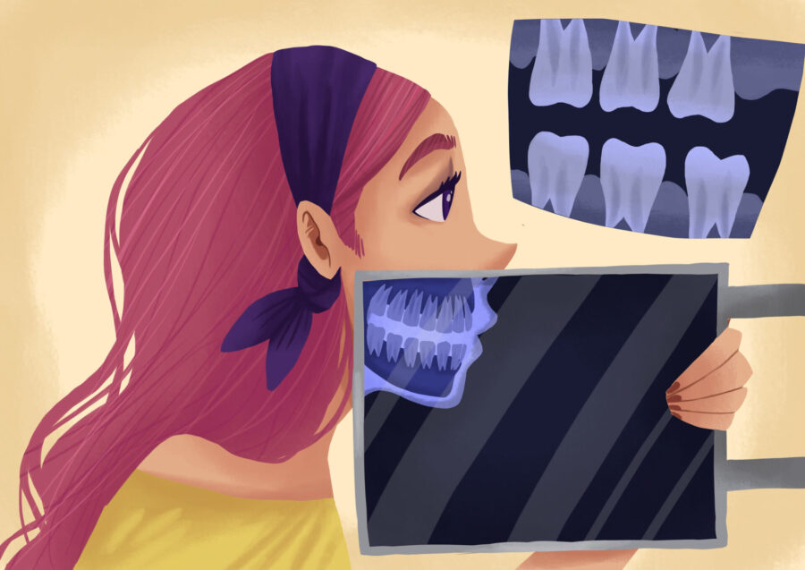
Dental X-rays are a common part of dental care, but it is normal to have questions about them. Most people know X-rays involve radiation, and that word can make even a routine dental visit sound more serious than it usually is.
The helpful thing to know is that routine dental checkup X-rays use a very small amount of radiation. They are also taken for a reason. Dental X-rays help dentists see between teeth, under old dental work, around the roots, and below the gumline, where problems are not always visible during a regular exam.
Compared with many everyday sources of radiation, the dose from dental X-rays is low. In fact, a routine set of dental X-rays does not come close to the radiation exposure many people get from flying on a plane.
At True Dental in Odenton, MD, Dr. Jai Shin and Dr. Julie Shin use dental X-rays when they are needed to check your teeth, roots, bone, and jaw more completely. The purpose is to get helpful information while keeping exposure low and appropriate for your dental needs.
Radiation Is Part of Daily Living
Radiation is not only something that happens during an X-ray. Everyone is exposed to small amounts of natural background radiation every day. It comes from the sun, soil, rocks, air, buildings, food, and even the natural materials around us.
On average, people in the United States receive about eight to ten microsieverts of natural background radiation per day. A microsievert is a unit used to measure radiation dose. The name sounds technical, but the comparison is simple: daily life already gives us small amounts of radiation exposure.
A routine dental checkup set is often in the same general range as part of a normal day of background exposure. In many cases, a few bitewing X-rays are around five microsieverts total. If one or two additional images are needed, the dose may be a bit higher, but it is still low.
That does not mean X-rays should be taken for no reason. It means the dose should be understood in context. When an X-ray can help find decay, infection, bone loss, or another dental issue early, the small exposure can give the dental team information that is hard to get any other way.
How Dental X-Rays Compare With Flying
Flying is one of the easiest ways to understand dental X-ray radiation because many people have been on a plane. When you fly, you are exposed to more cosmic radiation than you are on the ground. At cruising altitude, there is less atmosphere above you to block that radiation.
A routine set of dental checkup X-rays is much lower than many common flights. A short flight may expose you to several times more radiation than dental bitewing X-rays. A longer flight, such as a cross-country trip, can be far higher.
That does not mean flying is dangerous for most people. People fly for vacations, work trips, weddings, and family visits without giving radiation much thought. The comparison simply helps put the dental dose into perspective.
Dental X-rays are also focused on a small area. The image is aimed at the mouth, taken quickly, and used to answer a dental question. So, while the exposure is not zero, it is small and targeted.
If you have ever taken a flight without worrying about radiation from that trip, it may help to know that routine dental X-rays are generally much lower than the radiation from many flights.
What About Bananas and Food Comparisons?
Food can also help explain the scale. Bananas naturally contain potassium, and a tiny amount of that potassium is radioactive. One banana is often estimated at about 0.1 microsieverts of radiation exposure. That is an extremely tiny amount.
For comparison, many people receive about eight to ten microsieverts of natural background radiation during a normal day. So, one banana is only a tiny fraction of what your body is already exposed to from everyday life.
Bananas are safe to eat, and the radiation amount is far too small to be a concern. The comparison is helpful because it shows how tiny some radiation doses are when you put them next to normal daily exposure.
Dental X-rays are different from eating a banana. They are taken for a specific reason and aimed at a specific area. However, the bigger point is that a routine dental X-ray dose is small enough that everyday comparisons, like daily background exposure, bananas, and flying, help make the number easier to understand.
Why Dental X-Rays Have Clinical Value
A dental exam can show a lot. Dr. Jai Shin or Dr. Julie Shin can check your teeth, gums, bite, existing dental work, and visible signs of wear or damage. However, some dental problems start in places that cannot be seen by looking in the mouth.
Dental X-rays can help find cavities between teeth, decay under old fillings or crowns, bone loss from gum disease, infections near the roots, impacted teeth, and changes around the jaw. They can also help evaluate teeth before treatment such as extractions, root canals, crowns, implants, or dentures.
That clinical value is one reason X-rays are still such an important part of dentistry. They do not replace the exam, and the exam does not replace them. Together, they give the dentist a clearer picture of what is happening above and below the surface.
For example, a small cavity between two back teeth may not hurt. It may not show as a dark spot in the mirror. It may not feel different when you chew. A bitewing X-ray can show that cavity while it is still small, before it reaches the nerve or requires a larger repair.
X-rays can also show bone changes around teeth. This is especially helpful for patients with gum disease because bone loss can happen slowly. Looking at images over time helps the dentist see whether the condition is stable or getting worse.
They are also helpful when a tooth has been treated before. If there is a crown, filling, root canal, or bridge, the dentist may need an X-ray to see what is happening around the edges, roots, and surrounding bone. Without that image, part of the tooth’s story is hidden.
Dental X-rays have been studied and refined for many years. Guidelines for taking them are based on balancing diagnostic benefit with radiation safety. In practical terms, that means an X-ray should answer a real dental question, not just be taken because it is on a checklist.
At True Dental, your symptoms, dental history, cavity risk, gum health, exam findings, and treatment needs all help determine which images are worth taking.
Modern Dental X-Rays Use a Low Dose
Dental X-ray technology has changed a lot over the years. Many dental offices, like True Dental, now use digital sensors, which usually require less radiation than older film X-rays. Digital images also appear quickly, so the dentist can review them during the appointment.
Modern X-ray machines are designed to focus the beam on the specific area being imaged. That means the exposure is targeted. It is not spread broadly through the body.
A routine dental checkup set is often just a few small images. Bitewing X-rays show the crowns of the back teeth and the bone levels between them. If there is a specific tooth causing concern, a smaller image of that tooth may be added.
Even with several dental images, the dose remains low. It is very different from larger medical imaging tests that look at bigger areas of the body. Dental X-rays are focused on the mouth and taken for a short amount of time.
Dr. Jai Shin and Dr. Julie Shin can explain why a certain image is recommended and what they are looking for before moving forward.
If Dental X-Rays Are So Low, Why Does the Clinician Leave the Room?
This is a good question, and it comes up more often than people think. If the dose is so low for the patient, why does the dental team step behind a wall or leave the room to press the button?
The answer is repeated exposure. A patient may get dental X-rays once a year, or only when they are needed. A dental clinician may help take X-rays for several patients every day, week after week, year after year.
Even when each individual X-ray dose is very low, dental offices are designed to protect the team from repeated occupational exposure. Stepping behind a barrier, standing at a safe distance, or using shielded equipment helps keep that repeated exposure as low as possible.
Some offices may use handheld X-ray units, such as NOMAD-style devices, where the clinician stays in the room while using equipment designed with shielding. In other offices, the clinician may step out or stand behind a protected area. Both approaches are about safety and following the right protocol for the equipment being used.
For the patient, the exposure is brief and occasional. For the dental team, radiation safety is about what they do many times a day. That is why the room setup may look more cautious than the patient dose itself might suggest.
Why You May Not Need X-Rays at Every Visit
Not every dental appointment requires X-rays. The timing depends on your age, dental history, symptoms, risk for cavities, gum health, and whether treatment is being planned.
Someone with a history of frequent cavities, deep fillings, gum disease, or ongoing dental concerns may need X-rays more often than someone with very low cavity risk and stable dental health. A patient with tooth pain may need an image right away, even if their regular checkup X-rays are not due yet.
On the other hand, if your teeth and gums have been stable and there are no new symptoms, the dentist may recommend X-rays less often. The schedule should be based on what is clinically helpful, not on a one-size-fits-all rule.
During your visit, you can ask what the images are checking for or when your last dental X-rays were taken. The answer may be that the X-rays are needed to look for cavities between teeth, monitor bone levels, evaluate a painful tooth, or plan treatment.
If there is no current need, Dr. Jai Shin or Dr. Julie Shin may decide to wait. That kind of timing is part of using X-rays thoughtfully.
Dental X-Rays and Children
Parents often have extra questions when X-rays are recommended for children. That is fair. Children are still growing, and parents want to avoid anything unnecessary.
Dental X-rays for children are taken carefully and only when they can help answer a specific question. They may be used to check for cavities between baby teeth, monitor permanent teeth, evaluate dental injuries, or see whether teeth are developing in the right places.
Kids can get cavities between teeth just like adults, and those cavities are not always visible during an exam. Since baby teeth are smaller, decay can move through them more quickly. An X-ray can help catch a problem before it turns into pain or infection.
For orthodontic planning, X-rays may also show jaw development, missing teeth, impacted teeth, or teeth that do not have enough room to come in. That information can help families plan treatment at the right time.
At True Dental, Dr. Jai Shin and Dr. Julie Shin can explain why an X-ray is recommended for your child and how it fits into their dental care.
Dental X-Rays During Pregnancy
If you are pregnant or think you may be pregnant, tell the dental team before X-rays are taken. That allows the dentist to decide whether the image should be taken now, delayed, or adjusted based on your symptoms and treatment needs.
Many routine dental X-rays can wait if there is no urgent concern. However, if you have tooth pain, swelling, infection, or trauma, an X-ray may be needed to diagnose the problem and plan safe treatment. Dental infections should not be ignored during pregnancy.
Modern dental X-rays are focused on the mouth, not the abdomen, and the dose is very low. Still, the decision should be made thoughtfully. The dentist will weigh the benefit of the information against the timing and your specific situation.
The best step is to mention pregnancy before the appointment begins. Then Dr. Jai Shin or Dr. Julie Shin can talk with you about whether an X-ray is needed and why.
What About Lead Aprons and Thyroid Collars?
Many patients grew up having a heavy lead apron placed over them for dental X-rays. In some offices, aprons or thyroid collars may still be used depending on the image, equipment, patient, or office protocol.
The reason these protective coverings became familiar is simple: they were used to reduce exposure to areas outside the mouth. As dental X-ray technology has improved, the beam has become more focused and the dose has become much lower.
If a lead apron or thyroid collar is used, the team will place it before taking the image. If one is not used, you can ask why. In some cases, a collar may interfere with certain images, especially larger panoramic images, because it can block the view the dentist needs.
The team should be able to explain what type of X-ray is being taken, why it is needed, and what safety steps are used.
Types of Dental X-Rays You May Have
Different X-rays show different information. A bitewing X-ray is often used to check between the back teeth for cavities and to look at bone levels. These are the images where you bite down on a small tab or sensor.
A periapical X-ray shows the whole tooth, from crown to root tip. This type may be used when one tooth hurts, when the dentist is checking for infection, or when a root or surrounding bone needs a closer look.
A panoramic X-ray shows a wider view of the jaws, teeth, sinuses, and jaw joints. This image can be helpful for wisdom teeth, extractions, dentures, implants, orthodontic planning, or checking how teeth are positioned.
Some cases may require a three-dimensional image, often called a CBCT scan. This is not used for every patient. It may be recommended when more detailed information is needed for certain treatments, such as implants, complex extractions, or specific root canal concerns.
Each type of image has a purpose. Dr. Jai Shin or Dr. Julie Shin can explain which one is being recommended and what it will help show.
Balancing Safety and Good Dental Care
Dental X-rays are safe for most patients when they are used appropriately. The radiation dose is low, the images are targeted, and modern technology has made the process more efficient than it used to be.
At the same time, X-rays should still have a reason. They should help diagnose a problem, monitor a condition, plan treatment, or check areas that cannot be seen during an exam.
That balance is where good dental care comes in. Skipping needed X-rays can allow hidden problems to grow. Taking unnecessary X-rays does not help anyone. The right approach is thoughtful, based on your individual needs, and explained clearly.
If you have concerns about radiation, pregnancy, past medical treatment, or how often X-rays are being taken, bring those up. A short conversation can help you feel more comfortable with the plan.
Dental X-Rays at True Dental in Odenton, MD
A routine set of dental checkup X-rays uses a very small amount of radiation, often around the same scale as part of a normal day of background exposure. It does not come close to the exposure from many common plane flights, especially longer flights.
At True Dental in Odenton, MD, Dr. Jai Shin and Dr. Julie Shin use dental X-rays thoughtfully as part of your overall care. Call True Dental to schedule a visit and talk with the team about your dental health, your questions, and the safest way to get the information needed for your smile.
Image from Authority Dental under CC 2.0